Digital X-rays Therapy at Whiteboard Marketing
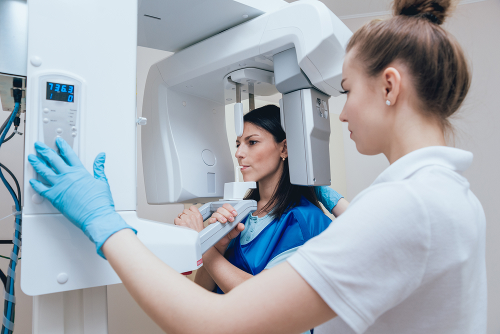
Dental x-rays capture in-depth, detailed images of a patient’s teeth and mouth that the naked eye cannot see. These x-rays are performed by our team of trained experts and they are typically considered to be one of the most common diagnostic tools in dentistry. These images allow [dr_name] to see a much clearer and more detailed view of our patient’s teeth, gums, and mouth which can reveal signs of infection, disease, trauma, bone loss, and many other issues.
The Benefits of Digital X-Rays
At Whiteboard Marketing, our goal is to provide the best, most accurate dental care possible for our patients. That’s why we are proud to offer the latest high-performing technology, such as digital x-rays, to better help treat our patients for all of their oral health care needs.
Depending on your discussion with [dr_name] regarding your unique dental situation and treatment plan, our team will typically perform digital x-rays on our patients periodically. Once these x-rays are taken, they are available instantly on our monitors and are stored electronically within your records for [dr_name] and the rest of our team to view.
These images aid [dr_name] in helping to detect and treat a number of oral health issues that may not be seen at first glance or without the use of x-rays at all. Compared to traditional x-rays, digital x-rays provide much clearer images and emit about 90% less radiation!
Intraoral X-Rays vs. Extraoral X-Rays
The most common type of digital x-rays that our practice uses are intraoral x-rays. A safe sensor is placed inside the mouth of the patient, which allows for a very highly-detailed image of the patient’s teeth, gums, and mouth. This allows [dr_name] to be able to view and detect a number of dental issues such as:
- Locating cavities
- Examining the roots and bones of the teeth and mouth
- View the overall health of the mouth, teeth, and gums
Extraoral x-rays are captured outside of the patient’s mouth. These types of x-rays are used to see the “bigger picture” of a person’s mouth and teeth and jaw structure. Extraoral x-rays help [dr_name] to precisely examine the following:
- Impacted teeth
- Tooth growth and development
- Facial bones
- Teeth and jaw connection
- Presence of cysts, tumors, and oral cancer
At Whiteboard Marketing, our goal is to provide the best dental care to our patients. Digital x-rays are an amazing technology that helps our team to accurately and efficiently detect, diagnose, and treat a variety of oral health issues. It is important to receive periodical dental x-rays at your appointment with us so our team can provide you with the best care! We encourage you to ask any questions you may have about x-rays by giving us a call today: [phone_number].
What Happens During the Digital X-rays Procedure?
Our dentists at Whiteboard Marketing ensure that every root canal procedure is as painless and efficient as possible. Here’s what you can expect:
- Local Anesthesia & Preparation—To ensure comfort, we begin by numbing the affected tooth with local anesthesia. Sedation options are available for anxious patients.
- Removal of Infected Material—We use specialized tools to remove infected material and bacteria from the pulp chamber and root canal system.
- Cleaning & Disinfection – The root of the tooth is thoroughly cleaned to eliminate any remaining infection.
- Filling & Sealing – The empty canals are filled with a biocompatible material and sealed to prevent reinfection.
- Temporary Filling & Final Restoration – A temporary filling protects the tooth until your general dentist restores it with a permanent crown or filling.
Most root canal treatments require one or two visits to complete. Following the procedure, you may experience mild discomfort, which can be managed with ibuprofen or other over-the-counter pain relievers.
Aftercare & Recovery
Proper aftercare is essential for a smooth recovery. To ensure the best results:
- Avoid chewing on the affected tooth until the final restoration is placed.
- Maintain good oral hygiene by brushing and flossing regularly.
- Take prescribed antibiotics if necessary to prevent reinfection.
- Schedule follow-up visits as recommended by your dentist.
A treated tooth can last a lifetime with proper care, helping you avoid extractions and maintain a natural, healthy smile.
Overflow
overflow content
Looking For Expert Digital X-rays Therapy in [practice_city]?
If you need expert care, Whiteboard Marketing is here to help. Our [ provide compassionate, high-quality [practice_type_adj] treatment options to protect and restore teeth. Call us at [phone_number] to schedule an appointment! Let us help you preserve the health and function of your teeth with advanced Digital X-rays therapy.